Call 408-257-5950 • 19000 Cox Avenue, Suite A • Saratoga, CA 95070
Why do I need a dental X-ray?

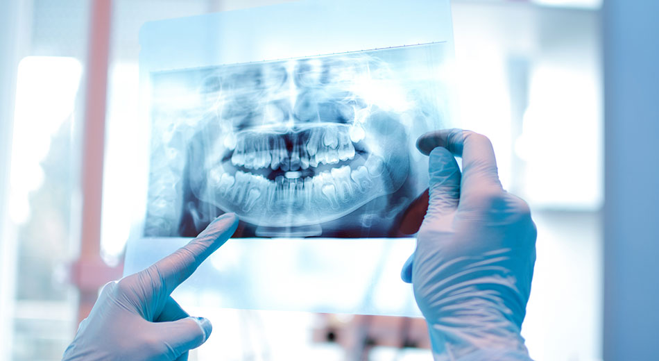
Dental X-rays are something you’ve undoubtedly seen before in your visits to the dentist throughout the years. But what is the significance of these X-rays, and why are we exposed to them? Dentists use dental radiographs (x-rays) to identify dental problems or damage that cannot be detected during a clinical oral exam.
How X-Rays Work
A small amount of electromagnetic radiation generates an X-ray image of your teeth, roots, gums, jaw, and facial bones during a dental x-ray. Dental radiographs, like other X-rays, use energy that can pass through less dense tissues but gets absorbed by solid objects.
The bright image on the X-ray is created when the dental Technician takes an exposure while the patient bites on a tab containing the film. This gives your dentist an internal view of your oral health.
Frequency of Dental X-rays
The frequency of dental X-rays should be decided on a case-by-case basis, according to the caries (tooth decay or cavity) risk assessment, according to the Food and Drug Administration (FDA) and American Dental Association.3 Some people are more likely to get tooth decay, so their dentist will recommend a different frequency of dental X-rays. Your caries risk fluctuates with time as well.
What They Detect
Dental X-rays can reveal various problems with your mouth, such as abnormalities that were not apparent on a visual examination. This is beneficial since the nature of your findings may influence how your dentist recommends treatments (for example, braces, implants, or wisdom tooth removal).
When your dentist takes X-rays of your mouth, they will look for:
- Position, size, and number of teeth
- Changes in the root canal
- Bone loss in the jaw or facial bones
- Bone fractures
- Tooth decay, including between teeth or under fillings
- Abscesses and cysts
- Impaction of teeth
- How the upper and lower teeth fit together
Dentists do a more extensive examination of the front teeth in children and young adults, looking for any missing (including number and size) teeth that haven’t yet developed. Adult teeth, wisdom teeth, or molars are all examples of this. They also inspect the jaw’s spacing to see how well the adult teeth will fit when they grow in.
How much radiation are you getting during an x-ray?
Dental x-rays are one of the safest and least harmful radiation treatments available. A typical check, which includes four bitewings, is approximately 0.005 mSv. That’s about the same amount of radiation you’d receive from the sun or a short flight of 1 or 2 hours daily. A panoramic dental x-ray delivers around twice as much radiation as a standard impression procedure (four bitewings).
Are x-rays safe for pregnant women?
If you must have an x-ray, your practitioner may suggest it be done after delivery. However, suppose x-rays are required because of a needed dental treatment that cannot be delayed. In that case, the American College of Radiology states that no single diagnostic x-ray has a radiation dose high enough to cause damage to an early embryo or fetus. Furthermore, dental x-rays show little risk of exposure to radiation outside of the teeth.
How often do you need an x-ray?
The frequency of dental x-rays varies depending on the patient’s oral health condition, age, the risk for disease, and any symptoms of oral disease. The American Dental Association has always maintained that dentists should only order dental x-rays for diagnosis or treatment when necessary.
Your dentist should also utilize the same techniques recommended by the ADA to minimize radiation exposure. Using abdominal shielding (such as protective aprons) is one method. The ADA also suggests that dentists utilize E or F speed film for standard x-rays, which reduces the amount of radiation needed for a good picture, or digital x-ray if available.
If you’re questioning the need for an x-ray, your dentist ordered, have a conversation with them about why they believe it’s necessary. Ultimately, you and your dentist must compare the benefits of getting the x-ray against the risk of being exposed to minimal radiation.
